
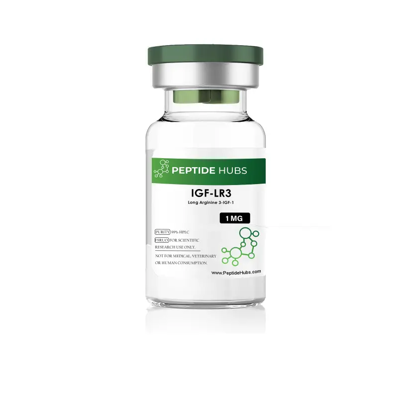
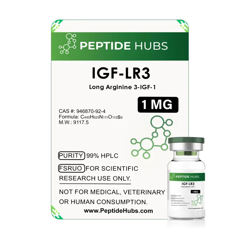
IGF-1 LR3
- USA MADE 2-7 days delivery
- International 5-20 days delivery
THIS PRODUCT IS INTEDED AS A RESEARCH CHEMICAL ONLY.
This designation allows the use of research chemicals strictly in vitro for testing and laboratory experimentation only. All products information available on this website is for educational purposes only. Bodily introduction of any kind into humans or animals is strictly forbidden by law. This product should only be handled by licensed, qualified professional. This product is not a drug, food or cosmetic and may not be misbranded, misused or mislabled as a drug food or cosmetic.
What is IGF-1 LR3?
Insulin-like Growth Factor 1 Long Arginine 3 (IGF-1 LR3) is a synthetic analog of human insulin-like growth factor 1, engineered to extend potency and duration of action in biological systems. Unlike the native IGF-1 molecule, which circulates for approximately 12–15 hours, IGF-1 LR3 remains bioactive for 20–30 hours, making it approximately three times more potent per administered dose. This extended pharmacological window derives from two key structural modifications: the substitution of arginine for glutamic acid at position 3 of the mature IGF-1 sequence, and the addition of a 13–amino acid N-terminal extension derived from porcine growth hormone.
The peptide retains full agonistic capacity at IGF-1 and insulin receptors, yet exhibits dramatically reduced binding affinity for insulin-like growth factor-binding proteins (IGFBPs)—natural inhibitors of wild-type IGF-1. This reduced binding to IGFBPs confers improved metabolic stability and allows the molecule to exert prolonged systemic effects. As an E. coli-derived recombinant protein with a molecular weight of approximately 9 kDa, IGF-1 LR3 has emerged as a preferred research tool in cell and tissue culture applications where sustained growth factor signaling is required.
IGF-1 LR3 Structure
The structure comprises eighty-three amino acids—a considerably more extensive assembly than the seventy resident within native IGF-1—arranged with the precision one might admire in an excellently ordered composition. The extended N-terminal sequence, MFPAMPLSSLFVN, precedes the mature IGF-1 coding region, concluding with AKA. Despite this extended architecture, the mature human IGF-1 core remains intact, ensuring facility for IGF-1 receptor engagement; however, the arginine substitution at position three, combined with the extended N-terminal housing, critically alters the protein's interaction with circulating IGFBPs.
This design yields a predicted molecular mass of 9 to 9.2 kilodaltons, verified by mass spectrometry analysis. The lyophilized formulation stabilizes the peptide in a shelf-stable state; upon reconstitution, the protein adopts its bioactive three-dimensional conformation. The modified structure preserves those critical disulfide bonds and secondary structures required for receptor binding whilst minimizing proteolytic degradation—a distinction of considerable advantage over native IGF-1 for long-duration research protocols.
IGF-1 LR3 Research
Cell Division
IGF-1 LR3 exerts a most vigorous and pronounced stimulus upon satellite cell proliferation and the accretion of myonuclei—those cellular processes foundational to tissue growth and the harmonious remodeling of muscular architecture. In serum-free culture models employing human breast cancer cell lines (MCF-7), IGF-1 LR3 displays an ED₅₀ of 0.3 to 1.5 nanograms per millilitre for cellular proliferation, indicating a potency and dose-dependency of the most gratifying order.
The peptide activates intracellular signaling cascades—principally the Ras-MAPK and phosphoinositide 3-kinase pathways—that flow downstream from IGF-1 receptor engagement, culminating in enhanced protein synthesis, cell cycle progression, and myogenic differentiation of a most satisfactory character. Research demonstrates with considerable clarity that IGF-1 LR3 stimulates both the proliferation and differentiation of skeletal muscle satellite cells, driving significant increases in muscle fiber size and myotube formation—effects which must be regarded as nothing short of felicitous.
The extended half-life confers a practical advantage of considerable moment: a sustained, low-level receptor activation persisting for twenty to thirty hours, producing cumulative cell signaling responses decidedly superior to equivalent bolus dosing of native IGF-1. In models of muscle regeneration and recovery—those most pressing concerns of the researcher engaged in tissue repair investigations—this sustained activation accelerates myogenic commitment and fusion, rendering IGF-1 LR3 a most valuable and indispensable tool for the understanding of satellite cell biology and the mechanisms by which tissue restoration is accomplished.
Fat Metabolism and Diabetes
IGF-1 LR3 modulates metabolic homeostasis through a dual engagement with cellular receptors: the simultaneous binding to both the IGF-1 receptor and the insulin receptor upon adipocytes, muscle tissue, and hepatic compartments. This multifaceted signaling increases glucose uptake into those tissues most sensitive to insulin's persuasion—particularly skeletal muscle, liver, and adipose compartments—via the enhanced translocation of GLUT-4, that most essential of glucose transporters.
The consequent decreases in circulating glucose levels trigger compensatory lipolytic cascades of considerable ingenuity: adipose tissue and hepatic stores undergo the methodical breakdown of triglycerides and glycogen, yielding a net shift toward energy expenditure and a reduction in adipose tissue mass of material significance. Research conducted in 3T3-L1 adipocytes confirms, with admirable consistency, that prolonged IGF-1 LR3 exposure enhances glucose uptake in a dose-dependent manner, with maximal effects evident at concentrations of twenty to one hundred nanomolar over incubations spanning twenty-four hours.
Clinical and preclinical data indicate with considerable assurance that IGF-1 LR3 reduces insulin requirements by approximately ten percent in hyperglycemic states—a finding of particular moment to those engaged in the investigation of type two diabetes management and the optimization of insulin sensitivity. The peptide's capacity to direct nutrients with admirable discretion—guiding glucose and amino acids toward muscle whilst increasing fatty acid oxidation—positions it as an instrument of the first importance for metabolic disorder modeling and intervention studies of the most demanding character.
Impairs Myostatin
Myostatin, that negative regulator of skeletal muscle growth, suppresses myogenic processes through a course of action executed via the TGF-β superfamily receptor signaling—specifically the ActRIIB pathway—and downstream Smad3-mediated transcription, effects of a most inhibitory character. IGF-1 LR3, however, exerts counteractive effects of considerable moment upon myostatin-driven muscle wasting, activating the IGF-1 receptor→PI3K/Akt axis, which antagonizes myostatin-induced myonuclei apoptosis and protein degradation with admirable efficacy.
In mouse models of Duchenne muscular dystrophy (DMD)—those lamentable conditions wherein muscle integrity is progressively compromised—IGF-1 LR3 and related IGF-1 derivatives demonstrate a capacity to mitigate myostatin-mediated pathology and preserve myocyte viability of the most encouraging order. The peptide functions, in considerable measure, by activating MyoD, that transcription factor commonly induced by exercise and tissue damage, thereby promoting muscle hypertrophy and regenerative myogenesis even in the presence of concurrent myostatin signaling.
Notably, the balance between IGF-1 signaling and myostatin levels, rather than muscle mass alone, emerges as critical to the maintenance of muscle strength and functional capacity—a distinction of no small importance to those engaged in the investigation of muscular physiology. Research suggests an inverse relationship of considerable elegance between constitutive IGF-1 overexpression and myostatin maturation, implying that sustained IGF-1 LR3 signaling may modulate myostatin proteolytic processing and bioavailability. This dynamic renders IGF-1 LR3 a powerful experimental agent of the first order for the dissection of myostatin biology and the evaluation of therapeutic strategies targeting muscle wasting diseases.
IGF-1 LR3 and Longevity Research
IGF-1 LR3 promotes tissue repair, cellular maintenance, and cellular stress resilience—attributes that position it as a most promising protective agent against age-related degeneration and cellular senescence. Preclinical studies conducted in mammalian models (cows, pigs, and mice) indicate, with gratifying consistency, that IGF-1 LR3 administration may attenuate those effects of cellular aging and age-associated pathology that commonly afflict the tissues—including dementia progression, muscle atrophy, kidney disease, and vascular dysfunction.
Glucocorticoid Signaling
Glucocorticoids exert strong catabolic effects upon skeletal muscle, impairing protein synthesis signaling and myogenic capacity. Concurrent IGF-1 signaling restores myogenic function by activating synthesis pathways that override glucocorticoid-imposed translational blockade. Combined glucocorticoid and IGF-1 therapy produces enhanced muscle recovery compared to monotherapy, a result of considerable encouragement to those engaged in tissue preservation research.
Feature Bullets
- Extended Half-Life (20–30 hours): Approximately three times more potent than native IGF-1 per equivalent dose; reduced IGFBP binding affinity ensures prolonged systemic availability and sustained receptor activation of considerable consequence.
- High Purity (99%): Recombinant E. coli-derived protein of the most exacting standards; meets stringent analytical requirements (SDS-PAGE, HPLC, mass spectrometry) satisfying the most demanding investigator.
- Robust Bioactivity: ED₅₀ of 0.3–1.5 ng/mL in serum-free cell proliferation assays; consistent myogenic and metabolic responses across replicate experiments of the most gratifying uniformity.
- Dual Receptor Engagement: Retains full agonistic activity at IGF-1 and insulin receptors with admirable fidelity; activates Ras-MAPK and PI3K/Akt signaling pathways for pleiotropic cell responses of considerable sophistication.
- Lyophilized Formulation: A most sensible and shelf-stable powder permitting long-term storage at −20 to −80 °C; convenient reconstitution workflow engineered for the efficient research laboratory.
- Myogenic and Metabolic Potency: Enhances satellite cell proliferation and myotube formation with notable vigor; increases glucose uptake and lipolysis with commendable efficiency; counteracts myostatin-mediated myonuclei apoptosis with demonstrated efficacy.
- Low Endotoxin Level (≤0.1 EU/μg): Reduced potential for spurious inflammatory responses in cell and tissue culture; admirably suitable for sensitive immunological assays and the most exacting experimental protocols.
Technical Specifications Table
|
Specification |
Value |
|
Product Name |
Recombinant Human IGF-1 LR3 (Long Arginine 3) |
|
Molecular Formula |
C₅₁H₇₈N₁₈O₁₄S₂ (calculated for 83-amino acid analogue) |
|
Molecular Weight |
~9–9.2 kDa |
|
Purity |
99% (SDS-PAGE and reverse-phase HPLC) |
|
Concentration (Stock) |
Typically supplied as 1 mg lyophilized powder per vial |
|
Form |
Lyophilized powder |
|
Source |
Recombinant E. coli expression |
|
Accession Number (UniProt) |
P05019 (human IGF-1 wild-type reference) |
|
Amino Acid Count |
83 amino acids (vs. 70 in native IGF-1) |
|
N-Terminal Extension |
MFPAMPLSSLFVN (13 additional amino acids) |
|
Key Substitution |
Arginine (Arg) at position 3 (vs. Glu in native IGF-1) |
|
Endotoxin Level |
≤0.1 EU/μg protein (LAL assay) |
|
Half-Life (In Vitro/Systemic) |
~20–30 hours |
|
Potency Relative to IGF-1 |
~3-fold higher per equivalent dose |
|
Storage Temperature |
−20 °C to −80 °C |
|
Stability Duration |
12 months from date of receipt |
|
Reconstitution Buffer |
Sterile PBS, water, or acetic acid (batch-specific) |
|
Formulation |
Lyophilized from filtered solution |
Storage Instructions
Temperature: −20 °C to −80 °C (a manual defrost freezer is most recommended, as automatic defrost cycles may prove injurious to the compound's structural integrity)
Stability: The compound shall remain stable for a period of twelve months from the date of receipt when stored at the recommended temperature, provided that proper protocols are faithfully observed.
Handling: One must exercise the utmost care to avoid repeated freeze-thaw cycles, those being most detrimental to the peptide's continued vitality. Should reconstituted product not be employed immediately, dilute the compound with concentrated bovine serum albumin to a final BSA concentration of 0.1 to 1.0 percent, thereby enhancing protein stability during storage. Centrifuge the vial with deliberate care before opening. Reconstitute by pipetting sterile diluent down the vial wall with measured composure; one must never resort to vortexing, an action most prejudicial to the molecule's structural integrity.
Reconstitution: Reconstitute in sterile phosphate-buffered saline, water, or acetic acid (the precise concentration shall be dependent upon one's specific experimental requirements) to achieve a stock concentration of at minimum 0.1 milligram per millilitre. Consult the batch-specific documentation provided for formulation-dependent reconstitution recommendations.
Endotoxin Level: ≤0.1 EU/μg protein, as measured by kinetic Limulus amebocyte lysate (LAL) analysis—a standard of purity and safety most rigorously maintained.

The reconstitution was crystal clear, a direct indicator of high purity and minimal filler. Peptide Hubs has become my most trusted source for high-affinity growth factor research.
The packaging was thermally insulated and discreet, ensuring the peptide’s structural integrity. I deducted one star only because of the precise handling required for such a potent 1mg vial, but the product quality itself is top-tier.
Related Products




Highly Purified Peptides: Trusted by Researchers Worldwide
Peptide Quality You Can't Find Elsewhere!!
| Peptide Hubs | Leading Competitor | |
|---|---|---|
| 200+ Peptide Variants |  |
 |
| USA-Manufactured Research Grade | |
|
| WHO/GMP & ISO 9001:2015 Certified | |
|
| Third-Party Tested for Purity | |
|
| Certificates of Analysis Provided | |
|
| Competitive Pricing Without Compromise | |
|
| Secure & Discreet Shipping | |
|
See Peptide Hubs in Action


Insights
Vesugen Peptide: Current Research on Aging and Vascular Biology

PE-22-28 Peptide: Exploring Its Mechanism and Scientific Potential

DSIP Peptide Research: Sleep Regulation, Evidence, and Safety
Cardiogen Peptide: Scientific Evidence and Potential Applications

99% Purity
Ensuring the highest standard with 99% purity.

Quality Ingredients
Crafted with purity and quality ingredients at the core.

Fast Shipping
Fast, reliable shipping straight to your door.

No TFA or Fillers
Nothing extra, just science: no TFA, no fillers.

Quality You Can Rely On — Guaranteed
At PeptideHubs, scientific integrity and product reliability are at the core of everything we do. Each compound is manufactured and tested under strict quality control protocols to ensure consistency, purity, and performance for research applications.
However, if for any reason our product does not meet your research expectations, we offer a 60-day money-back guarantee — providing a risk-free procurement experience for qualified researchers and institutions.
Your research deserves uncompromised quality — and we stand behind every order to ensure you receive.