
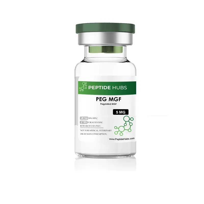
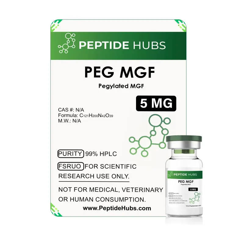
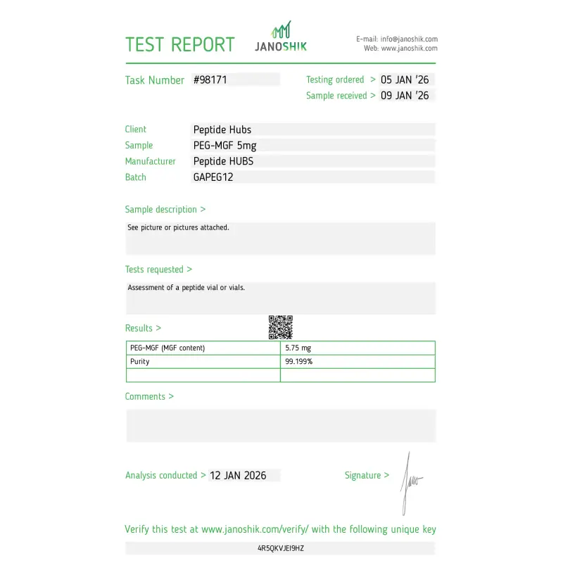
PEG MGF 5mg
- USA MADE 2-7 days delivery
- International 5-20 days delivery
THIS PRODUCT IS INTEDED AS A RESEARCH CHEMICAL ONLY.
This designation allows the use of research chemicals strictly in vitro for testing and laboratory experimentation only. All products information available on this website is for educational purposes only. Bodily introduction of any kind into humans or animals is strictly forbidden by law. This product should only be handled by licensed, qualified professional. This product is not a drug, food or cosmetic and may not be misbranded, misused or mislabled as a drug food or cosmetic.
What is PEG MGF?
PEG-MGF (pegylated mechano-growth factor) is a synthetic research peptide derived from the insulin-like growth factor 1 (IGF-1) gene. It represents the C-terminal E-domain of IGF-1Ec, also known as the E-peptide or mechano-growth factor. Mechanically, it is the last 24 amino acids of the naturally occurring IGF-IEc molecule.
Unlike native MGF, which possesses a half-life measured in minutes within the bloodstream, PEG-MGF undergoes pegylation—the covalent attachment of polyethylene glycol to the peptide backbone. This modification extends its circulatory stability and resistance to enzymatic degradation, transforming MGF into a long-acting research compound suitable for controlled in vitro and animal model studies.
Research indicates that MGF is released from muscle tissue in response to mechanical stress or injury, playing a localized role in cellular adaptation that differs mechanistically from systemic IGF-1. The pegylation process preserves the biological activity of the E-domain while substantially improving molecular half-life—extending stability from minutes to hours or longer depending on the research context.
PEG MGF Structure
Sequence: PEG-Suc-Tyr-Gln-Pro-Pro-Ser-Thr-Asn-Lys-Asn-Thr-Lys-Ser-Gln-Arg-Arg-Lys-Gly-Ser-Thr-Phe-Glu-Glu-Arg-Lys-Cys
Molecular Formula: C₁₂₁H₂₀₀N₄₂O₃₉
Molecular Weight: 2867.2 g/mol (an approximation dependent upon the PEG chain)
PubChem SID: 178101669
Synonyms: Pegylated MGF, PEG IGF-1 Ec, PEG myotrophin, PEG-MGF-E, PEG-MGF-Ct24E
The constitution of this substance is defined by the E-domain of IGF-1Ec, joined via a succinate linker to its PEG moiety. This arrangement confers a greater molecular weight and hydrodynamic radius, thereby discouraging rapid filtration by the kidneys and allowing the peptide to circulate with greater endurance. It should be noted that the PEG chains do not suffocate the peptide but rather form a negatively charged shield, protecting it from degradation while permitting it to engage with cellular receptors as propriety demands.
PEG MGF Research
PEG MGF and Skeletal Muscle
It has been observed that when skeletal muscle is subjected to the rigors of mechanical loading, it responds with a spirited upregulation of MGF mRNA. In studies concerning subjects of advanced years, resistance exercise was seen to increase MGF mRNA by some 163%, a figure that rose to 456% when accompanied by an elevation in growth hormone. Such findings suggest that MGF plays a principal role in the tissue's adaptation to exertion.
In murine models suffering from muscle contusion, the administration of MGF appeared to calm the inflammatory temperament of the tissue, reducing oxidative stress and the formation of fibrosis. Specifically, it was noted that the expression of disagreeable inflammatory mediators—such as TNF-α and IL-1β—was markedly diminished. Furthermore, the peptide appeared to discourage the excessive production of collagen types I and III, which are known to contribute to scarring.
Professor Goldspink and his associates have reported a 25% increase in muscle fiber diameter in active mice treated with MGF. However, the native peptide required localized administration to achieve such ends; the pegylated variant, happily, surmounts this limitation, allowing for systemic delivery without loss of efficacy.
PEG MGF Research in Heart Muscle Repair
Research emanating from the University of Illinois bioengineering department presents findings of exceptional interest. MGF inhibits apoptosis—that is, programmed cellular death—in cardiac myocytes subjected to hypoxic stress. When administered within eight hours of such stress, rats treated with MGF exhibited materially less cell death and greater recruitment of cardiac stem cells compared to controls.
The peptide appears to enhance the migration of mesenchymal stem cells through chemotactic mechanisms, drawing these multipotent cells toward sites of cardiac injury. Data suggests approximately thirty-five percent reduction in cardiomyocyte injury when MGF is administered during acute myocardial infarction, with improvements in hemodynamic function and reduction of pathologic cardiac remodeling.
Bone Repair and Growth
The restoration of bone requires a coordinated effort of osteoblast proliferation and mineral deposition. In studies involving rabbits with surgically induced defects, MGF was observed to accelerate the healing process substantially. Those subjects receiving the peptide exhibited a degree of recovery at four weeks which was not seen in the control group until six weeks had passed.
It is proposed that MGF influences the cell cycle, perhaps arresting osteoblasts in a phase most conducive to proliferation, whilst simultaneously activating the MAPK-Erk1/2 signaling pathway.
Protecting Cartilage
MGF enhances chondrocyte function and their migration from bone—their developmental origin—into cartilage tissue. Research indicates that MGF may delay cartilage degeneration, potentially through modulation of cellular stress response pathways.
The prolonged stability conferred by pegylation proves particularly advantageous for joint-space applications. A single intraarticular injection of PEG-MGF could theoretically exert protective effects across weeks or months, whereas unmodified MGF would remain active for mere minutes or hours.
Dental Applications
In vitro studies of human periodontal ligament cell cultures demonstrate that PEG-MGF enhances osteogenic differentiation and increases expression of matrix metalloproteinases MMP-1 and MMP-2. These proteases facilitate ligament remodeling and repair—those connective structures by which teeth are anchored to bone.
Research suggests potential applications in preserving dental tissues after injury or surgical reimplantation, offering an alternative to extraction or implantation.
Potential Neuroprotective Effects
Neurological investigations reveal that elevated MGF levels in the brain and central nervous system may slow age-related neuronal degeneration. Research in transgenic mice expressing elevated MGF demonstrated retention of cognitive function and peak cognitive performance into advanced age, with greatest benefits when MGF overexpression occurred early in life.
MGF treatment improved muscle weakness and reduced motor neuron loss in mouse models of amyotrophic lateral sclerosis. MGF is naturally expressed in the brain following hypoxic injury and is overexpressed in regions experiencing active neuronal regeneration, suggesting exogenous administration may reduce disease impact by preventing neuronal death despite ongoing pathology.
Feature Bullets
- Extended Plasma Stability: Pegylation extends MGF half-life from minutes to hours, enabling research applications of scope and durability
- 99% Purity by RP-HPLC: Pharmaceutical-grade purity ensures reliable, reproducible investigation of molecular effects
- Lyophilized Form: Freeze-dried powder simplifies storage, shipping, and handling whilst maintaining stability for extended periods at proper temperature
- Multi-Tissue Research Applications: Documented efficacy in skeletal muscle, cardiac muscle, bone, cartilage, periodontal, and neural tissue studies
- Mechanistic Clarity: IGF-1 receptor activation pathway and multiple downstream signaling mechanisms support controlled experimental design
- Consistent Molecular Profile: Standardized formula ensures batch-to-batch reproducibility essential for longitudinal investigation
Technical Specifications Table
|
Specification |
Value |
|
Peptide Name |
PEG-MGF (Pegylated Mechano-Growth Factor) |
|
Active Form |
24-amino acid E-domain of IGF-1Ec |
|
Concentration |
5 mg per vial |
|
Form |
Lyophilized (freeze-dried) white powder |
|
Purity |
99% (RP-HPLC) |
|
Molecular Formula |
C121H200N42O39 |
|
Molecular Weight |
2,867–2,948 Da |
|
Sequence |
PEG-Suc-Tyr-Gln-Pro-Pro-Ser-Thr-Asn-Lys-Asn-Thr-Lys-Ser-Gln-Arg-Arg-Lys-Gly-Ser-Thr-Phe-Glu-Glu-Arg-Lys-Cys |
|
Appearance |
White to off-white powder |
|
Storage (Unopened) |
–20°C; stable for 24 months from shipment |
|
Storage (Reconstituted) |
2–8°C for 1 month; –20 to –70°C for extended storage |
|
Recommended Diluent |
Sterile water or bacteriostatic water for injection |
|
Solubility |
Soluble in sterile aqueous solutions |
|
Suitable Applications |
Cell culture, animal research models, in vitro studies |
Storage Instructions
Lyophilized (Unreconstituted) Form:
The lyophilized powder should be kept at a temperature of -20°C, in a dry place, and strictly shielded from the intrusion of light. While the substance may endure room temperature for a season of three months, prudence dictates that sub-zero storage is superior for its preservation. For retention beyond a few months, a temperature of -80°C is advised.
Before the vial is unsealed, it must be allowed to reach room temperature within a desiccator, lest moisture condense upon the peptide—a circumstance that would surely compromise its stability. Once the necessary portion has been withdrawn, the vial should be resealed with haste and returned to the cold.
General Handling Precautions:
- Maintain the vessel in conditions cold, dry, and dark.
- One must avoid the repetition of freezing and thawing.
- Limit the substance’s exposure to the open air.
- Protect it from the direct gaze of light.

Shipping was prompt, though I would have liked a slightly more detailed breakdown of the PEG-linkage on the documentation. Overall, a very solid four-star product for serious metabolic labs.
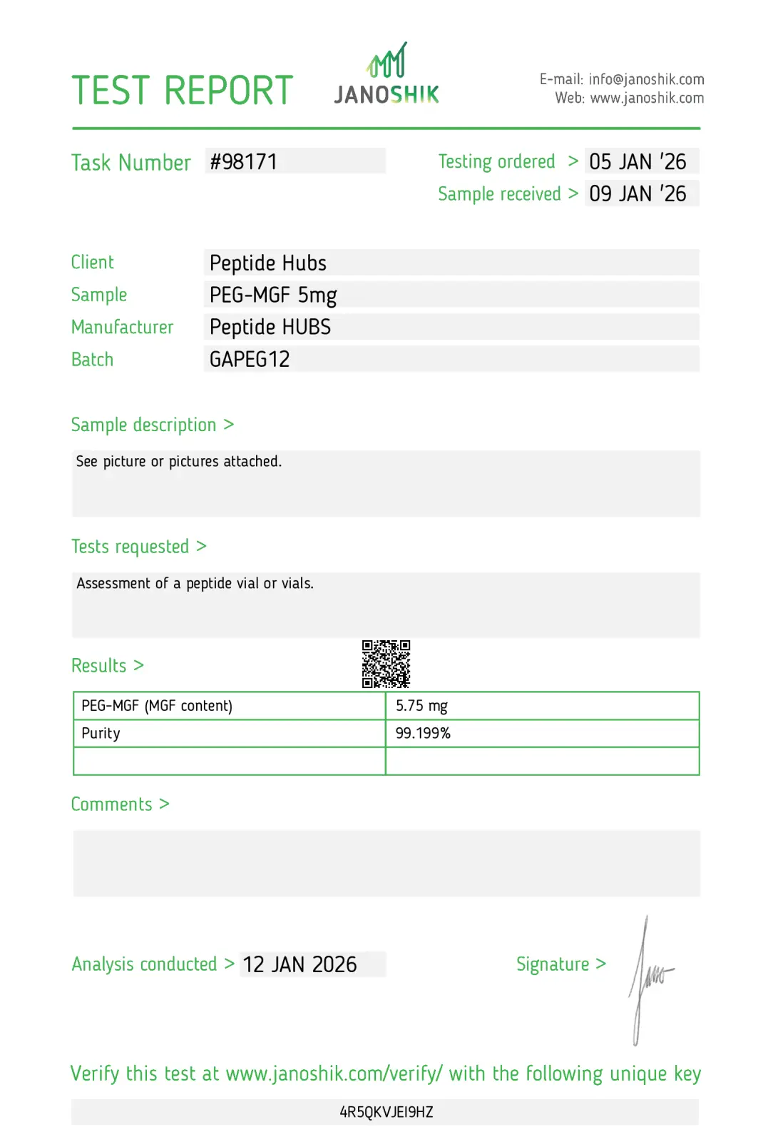
I was particularly impressed with the fast shipping and the discreet, professional packaging. For anyone researching tissue repair and lean mass preservation, this is a highly reliable source that matches its COA specifications perfectly.
The 5mg vial arrived vacuum-sealed with a uniform lyophilized cake. It is an indispensable tool for researchers focusing on long-term satellite cell recruitment.
Related Products




Highly Purified Peptides: Trusted by Researchers Worldwide
Peptide Quality You Can't Find Elsewhere!!
| Peptide Hubs | Leading Competitor | |
|---|---|---|
| 200+ Peptide Variants |  |
 |
| USA-Manufactured Research Grade | |
|
| WHO/GMP & ISO 9001:2015 Certified | |
|
| Third-Party Tested for Purity | |
|
| Certificates of Analysis Provided | |
|
| Competitive Pricing Without Compromise | |
|
| Secure & Discreet Shipping | |
|
See Peptide Hubs in Action


Insights

Vesugen Peptide: Current Research on Aging and Vascular Biology

PE-22-28 Peptide: Exploring Its Mechanism and Scientific Potential

DSIP Peptide Research: Sleep Regulation, Evidence, and Safety

Cardiogen Peptide: Scientific Evidence and Potential Applications

99% Purity
Ensuring the highest standard with 99% purity.

Quality Ingredients
Crafted with purity and quality ingredients at the core.

Fast Shipping
Fast, reliable shipping straight to your door.

No TFA or Fillers
Nothing extra, just science: no TFA, no fillers.

Quality You Can Rely On — Guaranteed
At PeptideHubs, scientific integrity and product reliability are at the core of everything we do. Each compound is manufactured and tested under strict quality control protocols to ensure consistency, purity, and performance for research applications.
However, if for any reason our product does not meet your research expectations, we offer a 60-day money-back guarantee — providing a risk-free procurement experience for qualified researchers and institutions.
Your research deserves uncompromised quality — and we stand behind every order to ensure you receive.