
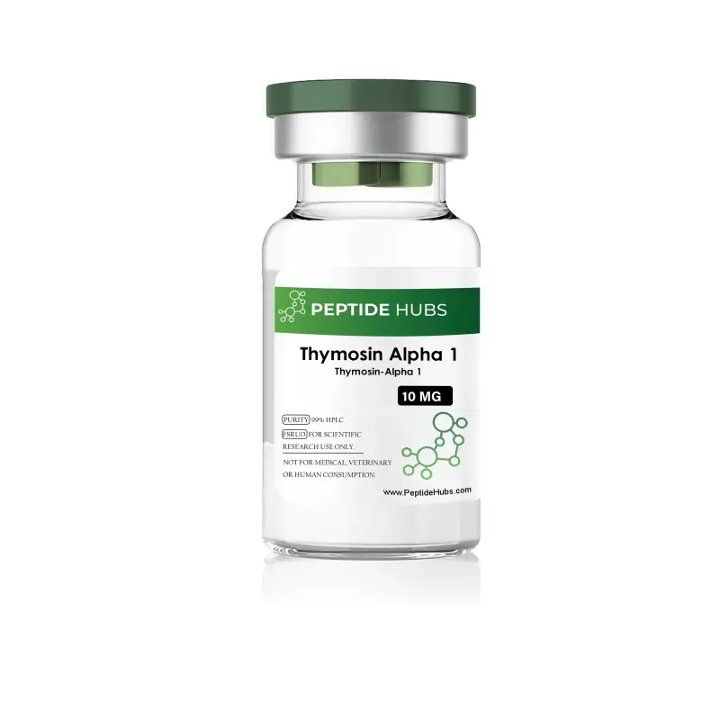
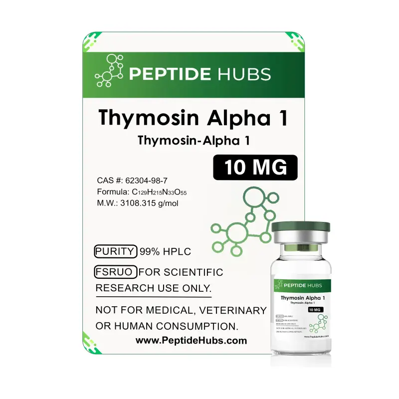
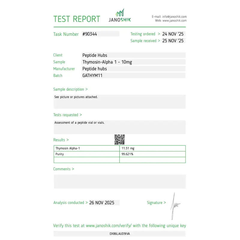
Thymosin Alpha-1 10mg
- USA MADE 2-7 days delivery
- International 5-20 days delivery
THIS PRODUCT IS INTEDED AS A RESEARCH CHEMICAL ONLY.
This designation allows the use of research chemicals strictly in vitro for testing and laboratory experimentation only. All products information available on this website is for educational purposes only. Bodily introduction of any kind into humans or animals is strictly forbidden by law. This product should only be handled by licensed, qualified professional. This product is not a drug, food or cosmetic and may not be misbranded, misused or mislabled as a drug food or cosmetic.
1. What is Thymosin Alpha-1?
Thymosin Alpha-1, designated TA1 or Tα1, is a naturally occurring peptide fragment first discovered in 1972 and extensively studied for its role in immunomodulation and physiological research. Known also by its synthetic designation, Thymalfasin, this bioactive polypeptide consists of 28 amino acids derived from a longer 113-amino-acid precursor termed prothymosin alpha. Originally isolated from thymus tissue, where it plays a central role in immune cell development, Thymosin Alpha-1 is now manufactured through solid-phase peptide synthesis for research purposes, ensuring consistent purity and quality.
Researchers hypothesize that Thymosin Alpha-1 functions as a regulator of adaptive and innate immune function by promoting T-cell maturation, enhancing natural killer cell activity, and modulating cytokine production. The peptide has been the subject of hundreds of clinical and laboratory investigations exploring its potential in diverse research domains, from immunological disorders to cellular regeneration and inflammatory responses.
2. Thymosin Alpha-1 Structure
A more methodical arrangement of twenty-eight amino acids could scarcely be imagined: Ser-Asp-Ala-Ala-Val-Asp-Thr-Ser-Ser-Glu-Ile-Thr-Thr-Lys-Asp-Leu-Lys-Glu-Lys-Lys-Glu-Val-Val-Glu-Glu-Ala-Glu-Asn. This linear peptide possesses, to the satisfaction of rigorous analytical science, a molecular weight of 3,108.31 g/mol, derived from its molecular formula: C₁₂₉H₂₁₅N₃₃O₅₅.
The structure itself demonstrates a most commendable capacity for purposeful engagement. The peptide exhibits particular affinity—one might observe with some satisfaction—for toll-like receptors, specifically TLR-2 and TLR-9, those sentinels positioned upon dendritic cells and antigen-presenting cells. Through such judicious binding, the molecule executes its influence upon immune responses with the precision one expects of well-conducted molecular machinery. Whether examined through mechanistic investigation or comparative structural inquiry, this three-dimensional arrangement permits Thymosin Alpha-1 to engage with cellular receptors and intracellular signaling pathways in a manner befitting serious scientific examination.
3. Thymosin Alpha-1 Research
3.1 Thymosin Alpha-1 Modulates the Immune System
The immunomodulatory character of this peptide proves most gratifying to those of us devoted to systematic investigation. Thymosin Alpha-1 demonstrates a decided propensity to enhance T-cell maturation and attendant function—a capacity that warrants consideration most deliberate.
Research of commendable rigor indicates the peptide possesses an inclination toward increasing major histocompatibility complex (MHC) class I molecules whilst simultaneously promoting cytokine production, those essential agents of adaptive immune response. Like a hostess of considerable skill managing her establishment with appropriate personnel, the peptide augments natural killer (NK) cell activity and fosters expression of phenotypic markers upon T cells—markers deemed critical for proper immune cell identification and purposeful function.
The evidence suggests, with gratifying consistency, that Tα1 increases expression of high-affinity interleukin-2 (IL-2) receptors upon cell surfaces, thereby promoting vigorous activation and proliferation of T lymphocytes. Clinical observations, accumulating with steadfast regularity, demonstrate that this peptide stimulates both T-helper and cytotoxic T-cell populations—those essential elements required for eliminating infected or aberrant cellular matter.
Furthermore, the peptide promotes differentiation of thymocytes and peripheral blood lymphocytes into mature immune cells with apparent facility. It encourages natural killer cell production, regulates alpha thrombin activity, and promotes cytokine-mediated inflammatory responses when circumstances deem such action appropriate. Research additionally explores its influence upon macrophage efficiency, revealing—to the satisfaction of the inquiring mind—broad immunological functionality of considerable scope and consequence.
3.2 Thymosin Alpha-1 Promotes Nerve Growth
One must acknowledge with some satisfaction that emerging investigations suggest Thymosin Alpha-1 may extend its beneficial influence to the nervous system's development and protection through mechanisms most ingeniously immune-mediated. Studies conducted with murine models of acknowledged rigor have demonstrated that the peptide potentially modulates genes responsible for neuronal growth and synapse development—work of considerable delicacy—whilst promoting with evident favor the expression of neurotrophic factors of distinction, including brain-derived neurotrophic factor (BDNF), nerve growth factor (NGF), and insulin-like growth factor-1 (IGF-1).
Concurrently, the peptide may reduce pro-inflammatory cytokines—chiefly interleukin-6 (IL-6) and tumor necrosis factor-alpha (TNF-α)—thereby fostering a neuroprotective immunological environment of marked salutary character. Research observations of the most gratifying sort include increased neuronal proliferation markers (BrdU), heightened neural stem cell markers (Nestin), enhanced neuronal progenitor markers (Tbr2), and mature neuron markers (NeuN), indicating enhanced neurogenesis proceeding with pleasing regularity.
Notably, and with evident significance, Thymosin Alpha-1 has been observed to impede lipopolysaccharide-induced impairment of hippocampal neurogenesis, positioning itself as both a neurogenic agent and neuroprotective factor against immune-mediated neurodevelopmental disruptions of the most troublesome character.
3.3 Thymosin Alpha-1 Fights Fungus
Dendritic cell maturation represents, in the estimation of experienced researchers, a matter of consequence most considerable. These cells, in their properly matured state, function as sentinels of discernment and propriety in fungal pathogen recognition and antifungal immune response.
Research demonstrates, with commendable clarity, that Thymosin Alpha-1 activates dendritic cells and promotes T-helper cell maturation in fungal infection models, particularly those involving Aspergillus fumigatus. The peptide enhances dendritic cell capacity to present fungal antigens to adaptive immune cells, rendering the immune system's response more efficient—a matter of evident satisfaction.
By promoting dendritic cell maturation through toll-like receptor signaling, Tα1 enhances both innate recognition and adaptive immune response generation to fungal threats. Given dendritic cells' strategic positioning within respiratory, cutaneous, and gastrointestinal tissues—those principal sites of pathogen entry—the peptide's demonstrated capacity to modulate these first-line defenders suggests relevance of genuine consequence across multiple infection scenarios.
3.4 Thymosin Alpha-1 and Hepatitis
Clinical investigations of considerable merit have explored Thymosin Alpha-1's potential in matters concerning hepatitis—a condition meriting the attention of serious practitioners. In subjects afflicted with hepatitis B and receiving the peptide with twice-weekly regularity, researchers documented virological response rates increased to the gratifying figure of 40.6%. For hepatitis C, subjects demonstrated improved outcomes when Thymosin Alpha-1 was administered in concert with interferon-alpha compounds—a partnership of considerable efficacy.
The proposed mechanism, advanced with proper scientific circumspection, involves modulation of cytokine profiles and enhancement of antiviral immune responses through toll-like receptor engagement upon dendritic cells.
3.5 Thymosin Alpha-1 and HIV
A randomized, open-label phase II clinical trial of documented rigor enrolled twenty subjects clinically stable and receiving highly active antiretroviral therapy (HAART), with thirteen recipients of Thymosin Alpha-1—administered twice weekly with becoming consistency—and seven others receiving placebo as counterbalance. Researchers monitored with meticulous attention CD4/CD8 cell counts, CD45 cell levels, and signal joint T-cell receptor circles (sjTREC) at two-week intervals throughout a twelve-week period.
Whilst CD4, CD8, and CD45 levels demonstrated no significant alteration in either group—a finding warranting neither surprise nor undue censure—sjTREC levels reportedly increased in peptide-treated subjects with gratifying consistency. Elevated sjTREC levels may indicate, scholars propose, enhanced thymic output and immune reconstitution of considerable consequence; however, this improvement did not correspond with increased total T-cell numbers or altered naive/memory T-cell phenotypes within the study's temporal constraints.
3.6 Thymosin Alpha-1 Research and Blood Pressure
Preliminary research has proposed that Thymosin Alpha-1 possesses dual antioxidant and angiotensin-converting enzyme (ACE) inhibitory properties. The peptide may function as a mixed ACE inhibitor, affecting both enzyme velocity and substrate affinity. ACE inhibition represents a recognized therapeutic strategy for blood pressure management, potentially relaxing blood vessels whilst delaying atherosclerosis progression and influencing renal function. Additionally, Thymosin Alpha-1 demonstrated antioxidant capacity in scavenging DPPH, ABTS, hydroxyl, and superoxide radicals, with reduced cellular reactive oxygen species (ROS) levels observed in neural astrocytoma cells. These findings remain preliminary; further detailed investigation is required.
3.7 Thymosin Alpha-1 Research and Cancer
Preclinical and clinical investigations have examined, with appropriate diligence, Thymosin Alpha-1's potential anti-proliferative activity across multiple cancer cell types—a pursuit of evident scientific consequence and human significance.
In lung cancer cell line studies (A549), the peptide demonstrated anti-proliferative effects at concentrations of twenty-four to forty-eight µg/mL, with apparent reduction in cell viability after twenty-four-hour exposure. These antiproliferative effects appeared accompanied, to the satisfaction of systematic observation, by enhanced antioxidant enzyme activities: catalase (at 12 µg/mL), superoxide dismutase (SOD), and glutathione peroxidase (GPx) at lower concentrations (6–12 µg/mL).
Thymosin Alpha-1 also reportedly inhibited A549 cell migration in concentration-dependent fashion and reduced cellular ROS production—consequences suggesting oxidative stress modulation within aberrant cells of considerable interest.
In hepatocellular carcinoma models, Tα1 exposure increased ROS levels in isolated leuko-monocytes while reducing ROS in HepG2 cancer cells, accompanied by delayed cell cycle progression and reduced cancer cell populations of gratifying extent. Research indicates dephosphorylation of Akt at Ser473 in cancer cells following Tα1 exposure, potentially explaining the reduced proliferation observed with such consistency.
Clinical studies document that long-acting Thymalfasin variants demonstrated enhanced tumor growth inhibition in breast and melanoma models compared to standard Tα1, with improved CD4/CD8 responses and elevated interferon-gamma and IL-2 levels—findings of considerable promise and merit. Additional investigations have examined Tα1 in colon, non-small cell lung, and hepatocellular carcinomas, yielding results of variable character requiring further investigation and refinement.
3.8 Thymosin Alpha-1 and Inflammatory Pain
Given documented anti-inflammatory potential in research models of respectable design, Thymosin Alpha-1 has been hypothesized by learned investigators to mitigate inflammatory pain perception through peripheral and central nervous system pathways of considerable sophistication. Murine research suggests with becoming evidence that the peptide acts directly at inflammation sites to reduce cytokine and inflammatory mediator production—chiefly tumor necrosis factor-alpha (TNF-alpha) and interleukin-1beta (IL-1beta)—potentially triggering pain signal modulation through mechanisms distinct from those employed by conventional anti-inflammatory analgesics.
Complete mechanistic elucidation remains, with proper scientific humility, under active investigation.
3.9 Thymosin Alpha-1 and Cystic Fibrosis
Cystic fibrosis, a condition of considerable gravity, presents complications that potentially include prolonged inflammation impairing mucous clearance and increasing infection rates, stemming from CFTR protein misfolding of the most troublesome character. Murine model research of acknowledged rigor has explored Thymosin Alpha-1's potential to reduce inflammation and improve CFTR protein function with gratifying preliminary results.
The peptide reportedly restored mucosal barrier integrity in inflamed gastrointestinal tracts of both normal and CF-specific murine models, as well as in metabolic syndrome models relevant to CF—observations of considerable consequence. These findings suggest Thymosin Alpha-1's protective potential may extend beyond pulmonary tissues to encompass the pancreas and liver, indicating broad-spectrum influence in mitigating CF-related organ complications of manifold character.
3.10 Damaged Teeth and Thymosin Alpha-1
Preliminary research of promising initial character indicates Thymosin Alpha-1 may improve repair of gingival and surrounding soft tissues following avulsion and replantation of permanent teeth with becoming efficiency, potentially promoting replanted tooth survival of considerable duration. Though initial findings command attention for their promise, comprehensive investigation remains, with proper scholarly caution, ongoing.
FEATURE BULLETS
- 99% Purity Assured: Lyophilized Thymosin Alpha-1 (10mg) verified through Certificate of Analysis and HPLC methodology of unimpeachable rigor
- Immunomodulatory Research Tool of Merit: A twenty-eight-amino acid peptide for investigating immune cell maturation and T-lymphocyte function with evident propriety
- Multi-System Research Applications: Studied across immunology, infectious disease, neurobiology, and oncology research contexts with consistency most gratifying
- Molecularly Well-Characterized: Molecular weight 3,108.31 g/mol (C₁₂₉H₂₁₅N₃₃O₅₅) with established amino acid sequence of precision exemplary
- TLR-Signaling Agent of Demonstrated Activity: Engages toll-like receptors (TLR-2, TLR-9) for investigating dendritic cell and immune cell activation with evident efficacy
- Extensive Research Documentation Provided: Supported by peer-reviewed clinical trials and preclinical in vitro/in vivo studies of considerable number and substance
- Laboratory-Grade Supply of Superior Provenance: Suitable for institutional research, academic studies, and biotech applications of all character and ambition
TECHNICAL SPECIFICATIONS TABLE
|
Specification |
Details |
|
Product Name |
Thymosin Alpha-1 |
|
Quantity |
10 mg |
|
Form |
Lyophilized Powder |
|
Purity |
>99% |
|
Molecular Formula |
C₁₂₉H₂₁₅N₃₃O₅₅ |
|
Molecular Weight |
3,108.31 g/mol |
|
Amino Acid Sequence |
Ser-Asp-Ala-Ala-Val-Asp-Thr-Ser-Ser-Glu-Ile-Thr-Thr-Lys-Asp-Leu-Lys-Glu-Lys-Lys-Glu-Val-Val-Glu-Glu-Ala-Glu-Asn |
|
Alternative Names |
TA1, Tα1, Thymalfasin |
|
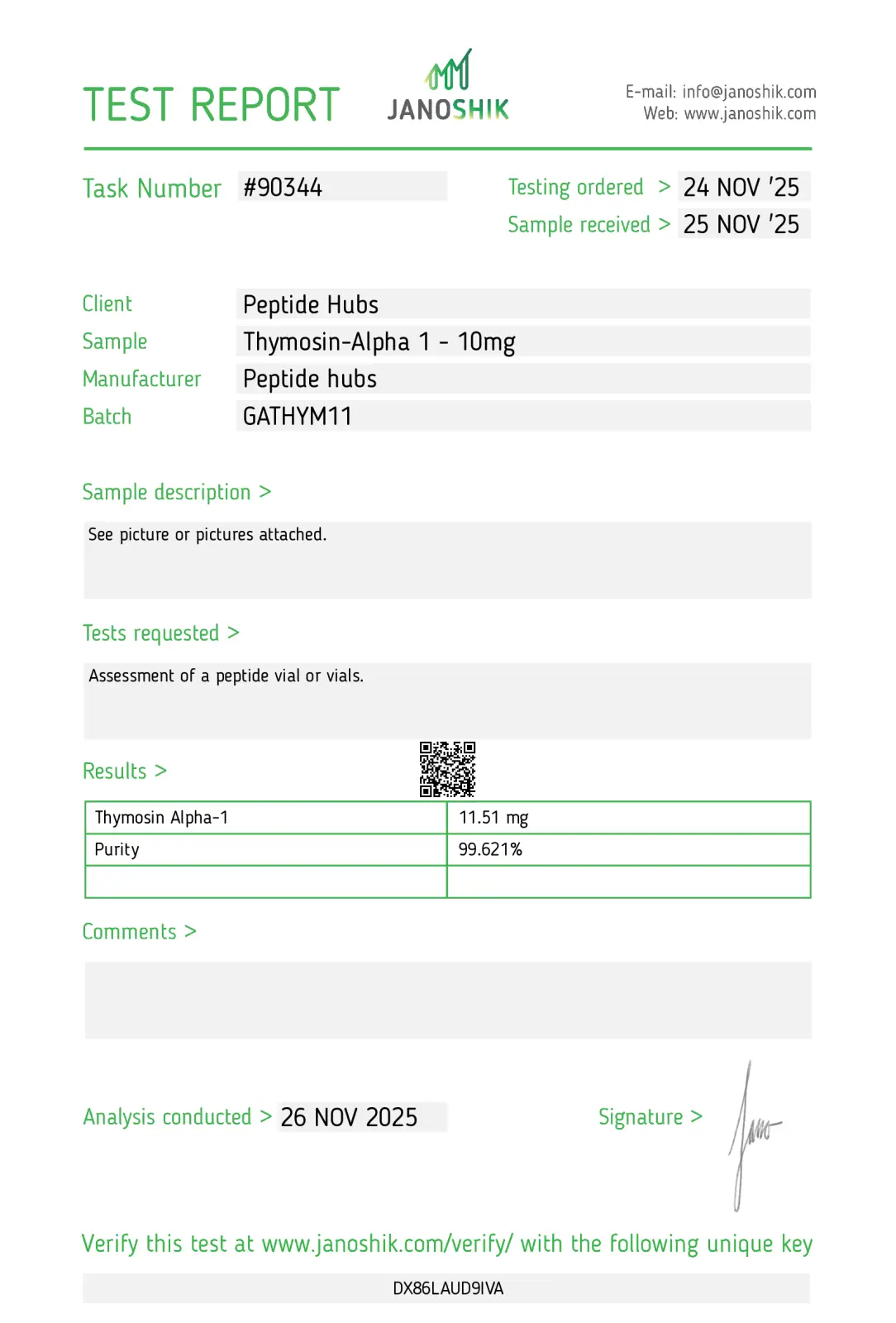
Certificate of Analysis |
Available upon request |
|
Quality Assurance |
HPLC, Mass Spectrometry, Certificate of Analysis provided |
Storage Instructions
Storage Conditions: Store lyophilized Thymosin Alpha-1 at -20°C (±5°C) in desiccated form for routine use. For long-term storage exceeding three months, maintain temperature below -18°C in a desiccated state.
Room Temperature Stability: Lyophilized powder remains stable for approximately three weeks at room temperature (20–25°C); however, prolonged ambient storage is not recommended.
Extended Storage: For durations exceeding three months, store at temperatures below -80°C in desiccated form, or maintain at -20°C with carrier protein supplementation (0.1% human serum albumin or bovine serum albumin).
Reconstituted Solution: Upon reconstitution with sterile water or saline, store reconstituted solutions at 4°C (refrigerated) for 2–7 days. For extended storage of reconstituted material, maintain at -20°C or below.
Critical Recommendations: Avoid repeated freeze-thaw cycles, which may compromise peptide integrity. Ensure proper desiccation prior to storage. Handle with care to prevent contamination.

Related Products






Highly Purified Peptides: Trusted by Researchers Worldwide
Peptide Quality You Can't Find Elsewhere!!
| Peptide Hubs | Leading Competitor | |
|---|---|---|
| 200+ Peptide Variants |  |
 |
| USA-Manufactured Research Grade | |
|
| WHO/GMP & ISO 9001:2015 Certified | |
|
| Third-Party Tested for Purity | |
|
| Certificates of Analysis Provided | |
|
| Competitive Pricing Without Compromise | |
|
| Secure & Discreet Shipping | |
|
See Peptide Hubs in Action


Insights
Bronchogen Side Effects and Safety: A Research Overview
GHK-Cu Safety Profile: Side Effects and Current Research

PEG MGF Side Effects: Safety, Tolerability, and Research Findings
PT-141 Safety Insights: Side Effects and Clinical Evidence

99% Purity
Ensuring the highest standard with 99% purity.

Quality Ingredients
Crafted with purity and quality ingredients at the core.

Fast Shipping
Fast, reliable shipping straight to your door.

No TFA or Fillers
Nothing extra, just science: no TFA, no fillers.

Quality You Can Rely On — Guaranteed
At PeptideHubs, scientific integrity and product reliability are at the core of everything we do. Each compound is manufactured and tested under strict quality control protocols to ensure consistency, purity, and performance for research applications.
However, if for any reason our product does not meet your research expectations, we offer a 60-day money-back guarantee — providing a risk-free procurement experience for qualified researchers and institutions.
Your research deserves uncompromised quality — and we stand behind every order to ensure you receive.